

3D Radiograph Lab
Choose an anatomy module, learn how its views are obtained, judge whether a film is technically adequate, and connect 2D landmarks to the structures that create them.
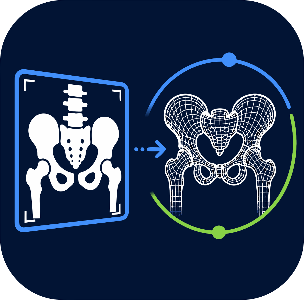
OrthoAnimate modules demonstrate concepts and support teaching. They are not validated patient-specific planning or clinical decision-support systems.
Start here
Select an anatomy tag, choose a projection, then rotate the 3D model while comparing it with the attenuation-based digitally reconstructed radiograph (DRR).
What you’ll learn 3 objectives
- Relate 2D projection landmarks to their 3D anatomical origins.
- Describe how common pelvis, hip, and lumbar-spine views are obtained.
- Recognise technical adequacy and common positioning errors.
This is a resident teaching model with editable assumptions and explicit limitations. It is not patient-specific and must not guide clinical decisions.
The left viewport shows an interactive CT-derived 3D anatomical model. The right viewport shows the corresponding attenuation-based simulated radiograph. Projection-specific acquisition guidance, quality criteria, purpose, landmarks, and measurements appear in the controls.
Read the projection, then reconstruct the anatomy
Module guidance will appear after the anatomy package loads.
Evidence & governance Assumptions · limitations · evidence · provenance
How to interpret this lab
Model assumptions
- The DRR integrates a simplified monochromatic attenuation estimate derived from Hounsfield units along parallel rays.
- The 3D surfaces are generated from physician-reviewed label volumes; the radiograph is generated from the aligned computed tomography (CT) volume.
- Hidden labelled structures are replaced with an approximate 40 HU soft-tissue attenuation rather than air.
- Hip positioning applies rigid femoral motion around the reviewed femoral-head centre. The optional skin surface and DRR soft tissue follow the same approximate deformation field; other labelled bones remain stationary.
Limitations
- The renderer does not model a clinical X-ray spectrum, scatter, beam hardening, detector response, image noise, or patient-specific exposure.
- Joint motion is an educational approximation without collision detection, dislocation constraints, muscle forces, or validated biomechanics.
- Soft-tissue deformation can leave artefacts and does not guarantee volume conservation. Folded regions use local best-fit estimates, not an extra limit on bone motion. The approximation requires further visual and clinical review.
- The current scan represents one individual anatomy and does not demonstrate normal population variation or every pathological pattern.
- Projected landmark and measurement guides are reviewed teaching annotations, not validated clinical measurements. Linear values use CT-space millimetres in this parallel-ray model and do not reproduce clinical radiographic magnification.
Evidence and references 11 selected sources
Projection selection and hip-evaluation teaching are informed by the following sources. View-specific content remains under active review while this module is in Preview.
- American College of Radiology, Society for Pediatric Radiology, and Society of Skeletal Radiology. Practice parameter for radiography of the extremities.
- Clohisy JC, Carlisle JC, Beaulé PE, et al. A systematic approach to the plain radiographic evaluation of the young adult hip. J Bone Joint Surg Am. 2008;90 Suppl 4:47–66. PubMed.
- Lim SJ, Park YS. Plain Radiography of the Hip: A Review of Radiographic Techniques and Image Features. Hip Pelvis. 2015;27(3):125–134. Full text.
- Yeap PM, Budak MJ. The pelvic radiograph: lines, arcs and stripes. Singapore Med J. 2021;62(7):333–340. Full text.
- American College of Radiology, American Society of Spine Radiology, Society for Pediatric Radiology, and Society of Skeletal Radiology. Practice parameter for spine radiography.
- Tazeabadi SA, Noroozi SG, Salehzadeh M, et al. Evaluation of Judet view radiographs accuracy in classification of acetabular fractures compared with three-dimensional computerized tomographic scan: a retrospective study. BMC Musculoskelet Disord. 2020;21:405. PubMed.
- Karkhur Y, Tiwari A, Maini L, Bansal V, Kakralia A. Radiological evaluation of pelvic inlet and outlet radiographic view in Indian population. J Clin Orthop Trauma. 2018;9(4):334–337. PubMed.
- Vrtovec T, Pernuš F, Likar B. A review of methods for quantitative evaluation of spinal curvature. Eur Spine J. 2009;18(5):593–607. Full text.
- Koslosky E, Gendelberg D. Classification in Brief: The Meyerding Classification System of Spondylolisthesis. Clin Orthop Relat Res. 2020;478(5):1125–1130. Full text.
- Chen X, Deng Q, Wang Q, et al. Image quality control in lumbar spine radiography using enhanced U-Net neural networks. Front Public Health. 2022;10:891766. Full text.
- Siddon RL. Fast calculation of the exact radiological path for a three-dimensional CT array. Med Phys. 1985;12(2):252–255. PubMed.
Attribution and provenance
The displayed CT volume, reviewed labels, generated meshes, processing history, source citation, and licence are defined by the selected dataset manifest. Dataset-specific attribution appears in the footer below.
Mesh and volume assets were prepared with OrthoAnimate Dataset Builder. The browser renderer uses a locally built subset of vtk.js; see Third-party notices.